
Nasal Tumours in Dogs and Cats
Nasal tumours are uncommon tumours in dogs and cats. They more commonly affect dogs than cats and are usually seen in older animals. The most common nasal tumour of dogs is carcinoma, which accounts for nearly two-thirds of all nasal tumours in this species. Carcinomas are malignant tumours that arise from the lining of the nose. Within this group the cancer may be described as an adenocarcinoma, squamous cell carcinoma and undifferentiated carcinoma, depending on the location within the lining of the nose from which the tumour arises. Sarcomas account for the bulk of the remaining nasal tumours. They usually arise from the cartilage, bone or connective tissue within the nose. The most common nasal tumours of cats are lymphoma (see ‘Lymphoma in cats’ handout) followed by carcinomas. Sarcomas are seen uncommonly in cats. Less commonly other tumours may be found in this location in both dogs and cats including melanoma and mast cell tumour. The following discussion is in relation to nasal carcinomas and sarcomas, for other tumours in this location treatment is often directed against the specific cancer. Like most tumours in animals and people, the cause of nasal tumours is unknown. However, long-nosed dogs and animals living in urban environments (with increased exposure to environmental pollutants) may have an increased risk of nasal tumour development.
Clinical signs
The most common presenting signs for animals with nasal tumours are sneezing, nasal discharge and nose bleeds. The discharge or bleeding will often start in one nostril and over time may affect both nostrils. There may be an initial, temporary improvement with antibiotics or anti-inflammatory drugs. These signs are not specific to cancer and may be seen with many diseases affecting the nasal cavity (i.e. fungal infection, foreign body, chronic inflammation). As the tumour progresses, further signs may be seen including facial deformity or swelling of the muzzle, loud raspy breathing, deviation or protrusion of an eye, and pain on opening the mouth. In some cases neurological signs such as seizures and behaviour changes may be seen in the few cases where the tumour extends into the brain. If the nasolacrimal duct is blocked by the tumour (a tube that drains tears from the eyes into the nasal cavity) increased or excessive tear production may be seen. Some more general signs associated with cancer include difficulty sleeping (often due to trouble breathing), decreased appetite and weight loss.
Diagnosis and staging
Diagnosis of a nasal tumour typically involves a complete physical examination, imaging of the affected area and a biopsy. A thorough inspection of the oral cavity is performed to screen for any changes (or bulging) of the hard palate, and oral lesions that may have extended into the nasal cavity (i.e. tooth root abscess). If the predominant clinical sign is nose bleeds we may recommend measuring the patient’s blood pressure and/or performing blood tests to assess the body’s ability to clot the blood properly (coagulation tests). Following this, imaging of the area is performed which may include radiographs, MRI, CT scan and endoscopy. Often one or a combination of these imaging techniques is used. Radiographs of the nasal passages may show destruction of the normal bony structures (turbinates) of the nose and/or a soft tissue mass. Normal radiographs do not rule out a tumour as they are the least sensitive of the imaging methods. More advanced imaging (MRI/CT) provides greater detail and information on the size of the tumour and infiltration of surrounding tissues.
Endoscopy involves passing a long, usually flexible tube (endoscope) with a camera at one end into the nasal cavity. This is passed one of two ways, either via the nostril or via the mouth, to view the front or back of the nasal cavity respectively. This allows visualisation of most areas of the nasal cavity, however because there are many passages of varying size, it is not possible to enter all of these passages and to view the entire nasal cavity. Following completion of the imaging tests biopsies are obtained. If the mass can be visualised with the endoscope biopsies can be guided towards the tumour. If the mass cannot be visualised biopsies are often collected ‘blind’. In this case the biopsy instrument is directed to the area of the tumour (based on advanced imaging). A biopsy involves taking a small sample of tissue from within the tumour. Most of the time a biopsy will provide a diagnosis as to the cause of the nasal signs and if it is cancer, the type of cancer involved (i.e. carcinoma versus sarcoma). In some cases the biopsy is non diagnostic or do not accurately represent the disease process. Repeat biopsies may be necessary if clinical signs persist.
Once the diagnosis of nasal cancer is made the patient is assessed to establish if there is evidence of cancer spread to other areas of the body (metastases). This is called staging. Generally nasal tumours are locally invasive and they infrequently metastasise. They tend to do so late in the disease. Staging for nasal tumours may include palpation and fine needle aspiration of local lymph nodes and chest radiographs. Blood tests (a complete blood count and biochemistry) and urinalysis are also performed to establish the general health of the patient and organ function prior to treatment.
Treatment
Treatment options for nasal tumours include surgery, chemotherapy and radiation. Radiation therapy involves the local application of a form of radiation (megavoltage) onto the area where the tumour is located. This is a local treatment only and therefore it is important to stage the patient prior to radiation to screen for disease elsewhere. Unfortunately, access to radiation in Victoria is limited and this would require travel to Queensland. Chemotherapy is the next best option and is indicated in all cases with evidence of metastases (spread of the cancer). The most commonly used chemotherapy drugs for nasal tumours are carboplatin and doxorubicin and we typically use them in combination with a non-steroidal anti-inflammatory drug registered in humans called piroxicam. Chemotherapy is generally well tolerated in animals. Please see ‘Chemotherapy in animals’ handout. Surgery may be considered if the mass is very small and well demarcated (i.e. not infiltrative), however this is rarely the case and surgery is often unrewarding. Orthovoltage radiation can be used but only after surgical removal of the bulk of the tumour. For some the above treatments are not feasible and palliation with piroxicam alone may decrease clinical signs for a period of time. However, in some cases there is no response and clinical signs persist.
Prognosis
Most dogs treated with radiation or chemotherapy experience an improvement in their clinical signs (less sneezing and/or bleeding) and some experience significant tumour shrinkage for a long period of time. The average survival time of patients that do not undergo treatment is three to six months. The average survival time for patients treated with radiation is 12–18 months, and some other studies have reported even longer averages (up to 20 months). There is less information available for chemotherapy, however one study of eight dogs showed an average survival time of 12–18 months, which is comparable to radiation. Surgery alone may result in a cure in rare cases where the tumour is small and contained to one area which is rare in the nose. However, usually some tumour will be left behind, and surgery without adjunctive treatment (radiation, chemotherapy) does not tend to improve survival times. Most animals with a nasal tumour eventually succumb to signs relating to the local extent of the tumour rather than from the spread of the disease.
Follow up
Following the completion of chemotherapy we recommend periodic rechecks to screen for recurrence or metastasis. Repeat imaging of the area is ideal but is often cost prohibitive and we often monitor patients based on clinical signs. Rechecks are typically recommended one month after finishing chemotherapy and then every three months thereafter. Early detection of recurrence or metastases is often beneficial and allows prompt management or treatment.
Related reading:
Information provided by Melbourne Veterinary Specialist Centre (MVSC), part of the AREN network.
Looking for additional information info on common pet illnesses and diagnoses?
For referring vets, please use our online referral form to submit a case enquiry.
You might also be interested in

Allergies In Dogs | Causes, Symptoms & Treatment
Discover the causes, symptoms, & treatments for allergies in dogs, from itchy skin to food sensitivities. Learn how to keep your pet comfortable & healthy.
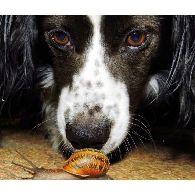
Garden Snails & Slugs Pose Life-Threatening Risks To Dogs & Humans
Learn about garden snails in your home, if they are poisonous to dogs & humans, symptoms and treatments from expert Veterinarians.

Arthritis In Dogs | Causes, Symptoms & Treatment
Learn about arthritis in dogs, including its causes, symptoms, and diagnosis. Discover ways to manage & improve your dog’s comfort and quality of life.
