
Canine Mast Cell Tumours
Mast cell tumours (MCTs) are the most common skin cancer in dogs. They are less common in cats and extremely rare in humans. Mast cells are normally involved in the body’s response to allergens and inflammation. They can be found in any tissue in the body and small numbers circulate in the bloodstream. A MCT occurs when there is uncontrolled growth of cancerous mast cells. They are considered malignant, meaning they have the ability to spread to other areas of the body and invade into the local area. Although MCTs occur most often in the skin, they can occur in any area of the body, particularly the liver and spleen. Mast cells contain substances (such as histamine) that can be released into the bloodstream and cause adverse effects elsewhere. These include allergic or anaphylactic reactions, stomach or intestinal ulcers and bleeding. Any dog can develop a MCT but certain breeds are predisposed. Mast cell tumours are more common in middle-aged to older animals, and can also be found in young animals.
Clinical signs
Mast cell tumours in the skin can look and feel like anything (they are sometimes referred to as the ‘great pretenders’). It is therefore important to have all new skin masses assessed by taking a fine needle aspirate and checking it under a microscope. Mast cell tumours that occur in locations other than the skin may have general non-specific clinical signs (such as weight loss, inappetence, lethargy) or signs relevant to the area affected (such as diarrhoea for stomach and intestinal MCT).
Diagnosis grading and staging
The diagnosis of MCTs involves taking a fine needle aspirate or biopsy. A fine needle aspirate is a simple test done with a small needle and syringe. The sample is usually sufficient to diagnose a MCT but does not give us any information on the grade (i.e. high versus low grade). A biopsy involves taking a larger sample often with sedation or local anaesthesia, but sometimes requires a general anaesthesia. The large sample provides information on the grade of the tumour and the rate at which it is dividing—called the mitotic index. These are important to help predict how the tumour will behave, which influences treatment and prognosis.
There are three grades of mast cell tumours:
- Grade 1 are the least aggressive and only occasionally spread to other parts of the body.
- Grade 2 are more aggressive as they extend more deeply into surrounding tissues. They may metastasise (or spread) to the local lymph nodes, spleen, liver or bone marrow.
- Grade 3 carry a poorer prognosis as the tumour tends to invade skin and underlying tissue and they have a high rate of metastases.
Recent studies have shown that the number of dividing cells seen on biopsy (mitotic index) is a very important indicator of how the tumour will behave. Staging involves further tests to determine if the cancer has spread to internal organs (these are called ‘metastases’). Staging for a MCT may include an abdominal ultrasound, chest radiographs, fine needle aspiration or biopsy of local lymph nodes, and possibly a bone marrow aspirate. It should also involve a careful examination of the skin over the entire body to screen for other skin masses. It is not uncommon for more than one MCT to be found. We also perform blood and urine tests to assess the health of the patient prior to treatment and to screen for mast cells circulating in the bloodstream (this is uncommon).
Treatment
Treatment options for MCTs include surgery, chemotherapy, radiation therapy and symptomatic treatment. Surgery is usually the best treatment option. It involves removing the tumour with a wide margin of normal tissue both around it and underneath it to increase the likelihood of removing the entire tumour. The margin of normal tissue that is taken is determined by the grade and location of the tumour (which is why having a biopsy before surgery can be useful). Because these tumours can be invasive, even when a large margin is taken, sometimes tumour cells can be left behind. A pathologist will endeavour to determine whether the entire tumour was removed. If there are tumour cells remaining additional treatment is sometimes necessary, which may include further surgery, chemotherapy, or radiation.
Surgery is not always possible, particularly for a tumour that is too large or located in an area that is not amenable to surgery either within the skin or elsewhere in the body (i.e. liver, gastrointestinal tract). Sometimes multiple tumours develop simultaneously with such rapid frequency that surgery will not be effective. Chemotherapy is used if the cancer cannot be removed with surgery, has a high risk of spreading (i.e. high grade or mitotic index), has already metastasised or spread, or if some residual cancer remains after surgery. The most commonly used chemotherapy drugs for MCTs are vinblastine (injection) and CCNU (tablet), and we use a combination of these. Approximately 80% of patients with MCTs will have some response to the chemotherapy. Chemotherapy is generally well tolerated in animals (for more information please see ‘Chemotherapy in animals’ handout). Radiation therapy can also be used to treat mast cell tumours, especially when surgery is not possible or there is residual cancer left after surgery. Radiation is a local treatment only and does not treat metastatic disease. It must be combined with chemotherapy to treat disease elsewhere in the body in high risk tumours. Unfortunately, access to radiation in Victoria is limited—this would currently require travel to Queensland.
We may also prescribe additional medication to treat or prevent MCT related side effects. These include an antihistamine (such as Zyrtec) and an antacid (for gastrointestinal ulceration or bleeding). It is important to monitor patients for evidence of ulcers or bleeding in the gastrointestinal tract. This may manifest as very dark or black stools, vomiting (with or without blood) or loss of appetite.
Prognosis
The prognosis for MCTs depends on many factors including the grade and mitotic index, the location of the tumour, the presence or absence of spread, and whether or not the tumour is new or a recurrent tumour (i.e. regrowth of tumour previously removed). Many are successfully treated (or cured) if they are low grade and there is no evidence of spread at the start of treatment. For those that have already spread, have recurred, cannot be removed or that occur in locations other than the skin (i.e. liver or spleen) the prognosis is less favourable. The goal of treatment in these cases is often to shrink the tumours and maintain a good quality of life for as long as possible. The grade of the tumour can be used to predict approximate survival times:
- Grade I: approximately 90% of patients live more than 12 months after surgery alone.
- Grade II: approximately 80% of patients live more than 12 months after surgery alone.
- Grade III: approximately <20% live more than 12 months after surgery alone but with the addition of chemotherapy approximately 70% live more than 12 months.
The mitotic index (or rate at which the cells are dividing) can also help to predict survival times, particularly for the intermediate grade (II) in which there can be a lot of variability. For Grade II tumours if the mitotic rate is high (greater than 5) the average survival time is five months, compared to 70 months if the rate low (less than 5).
Follow up
We recommend follow up rechecks one month after chemotherapy then every three months. This may involve checking the site of the tumour as well as local lymph nodes and may involve further staging tests (for example abdominal ultrasound). Up to 50% of dogs with previously diagnosed MCTs develop another new MCT at some stage in their life at a different location, usually in the skin. Therefore it is important to continually monitor the skin for new masses. If new masses arise it is important that they are evaluated with a fine needle aspirate, as early diagnosis and treatment will improve the outcome.
Related reading:
Information provided by Melbourne Veterinary Specialist Centre (MVSC), part of the AREN network.
Looking for additional information info on common pet illnesses and diagnoses?
For referring vets, please use our online referral form to submit a case enquiry.
You might also be interested in
Anal Sac Adenocarcinoma in Dogs
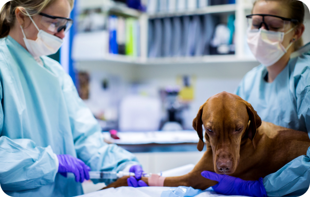
Find out the signs, diagnosis and treatment options for anal sac adenocarcinoma in dogs, from the specialists at MVSC.
Nasal Tumours in Dogs and Cats
Learn the causes, symptoms, diagnosis and treatment options for nasal tumours in dogs and cats, from the specialists at MVSC.

Diseases of the Nose in Dogs
Learn about common nose diseases in dogs, including DLE, pemphigus, solar dermatitis and mucocutaneous pyoderma, from MVSC.
