
A Practical Guide to Veterinary Surgical Oncology: From Pre-Op Staging to Post-Op Margins
By Dr Charles Kuntz DVM, MS, MACVSc, Diplomate ACVS, ACVS Founding Fellow of Surgical Oncology, Registered Specialist of Small Animal Surgery, Senior Staff Surgeon
When it comes to cancer surgery in pets, few things linger longer in a vet’s mind than a local tumour recurrence that could have been avoided. Incomplete surgical margins don't just compromise outcomes—they lead to repeat surgeries, emotional stress for owners, and professional frustration.
This article outlines the essential principles of surgical oncology to help ensure your first surgery is your best shot—because in cancer treatment, wide margins can mean the difference between cure and recurrence.
The Importance of a Realistic, Integrated Approach to Cancer in Pets
Cancer in dogs and cats can be overwhelming—but it isn’t always terminal. An optimistic yet realistic mindset is critical. Collaborating with pet owners to establish treatment goals, while balancing tumour biology with available therapies (surgical, medical, and radiation oncology), leads to the best outcomes.
While multimodal treatment is often required, surgery remains the cornerstone for diagnosing and definitively treating solid tumours.
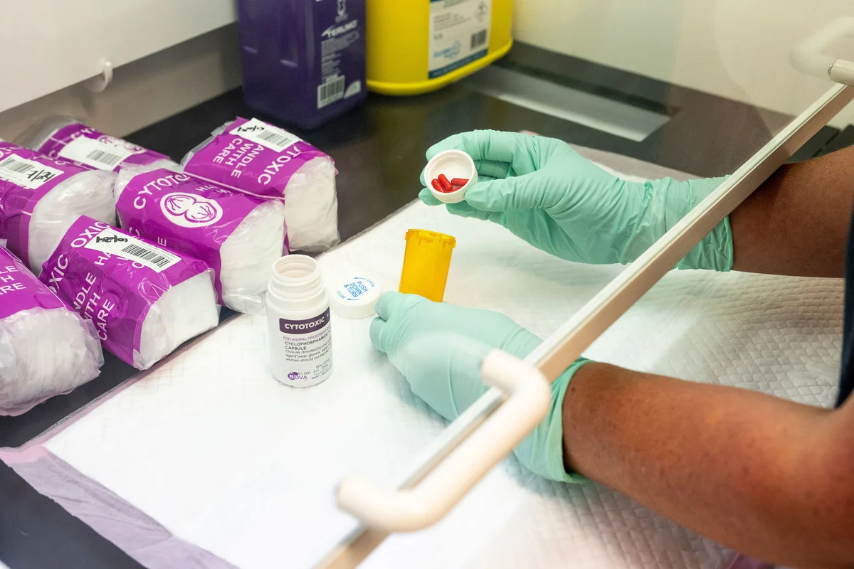
1. Pre-Operative Assessment: Look Beyond Chronological Age, start with a comprehensive evaluation of the pet’s:
Overall health and signalment
Concurrent diseases or conditions
Chronologic age alone is a poor predictor of surgical resilience. Physiological status matters most. Stabilising comorbidities—even if unrelated to the tumour—can significantly reduce perioperative risk.
2. Rigorous Cancer Staging: Go Beyond the Surface
Accurate staging is non-negotiable. It informs:
Prognosis
Treatment strategy
Owner communication
Use the WHO TNM system to assess local, regional, and distant disease. A baseline diagnostic workup should include:
Diagnostic biopsy
3-view thoracic radiographs
Fine-needle aspirates (FNA) of regional lymph nodes
Note: Palpation is not sufficient. Depending on tumour type, add abdominal ultrasound or
CT imaging for full characterisation.
3. Biopsy: Do It Right the First Time
A properly planned biopsy provides critical insights into:
Tumour type and grade
Biological behaviour
Key biopsy principles:
Choose tools that avoid cell damage
Orient the incision so the tract can be excised en bloc with the tumour
Ensure margins remain uncompromised
Skipping biopsy planning risks misdiagnosis—and compromises definitive treatment.
4. Pathology Submission: More Than Just Sending a Sample
Fix tissues in 10% buffered formalin (minimum 1:10 tissue-to-formalin ratio). Always provide detailed clinical history to the pathologist.
On receiving results:
Review tumour type, grade, and margin status carefully
Don’t hesitate to query anomalies, request re-sectioning, or seek a second opinion
5. Define the Surgical Goal: Diagnosis vs. Cure
Each oncology case must have a clearly defined objective - typically diagnosis, cure, or both. For localised tumours, first surgery offers the best chance of cure.
6. Surgical Dose: Err on the Side of Aggression
Select your approach - marginal, wide, or radical - based on:
Tumour behaviour
Prognosis
Comorbidities
Client expectations
Adjunct therapies
Common mistake: Under-dosing.
For malignant masses, a controlled open wound is preferable to leaving tumour cells behind.
Rule of thumb:
Wide margins of 2–3 cm in all directions, plus fascial planes for depth, are standard for tumours prone to local recurrence.
7. Patient and Site Prep: Think Ahead
Prepare the site with wide clipping to allow for extended margins. Address any paraneoplastic syndromes pre-op. If adjuvant therapy (e.g., radiation or reconstruction) is likely, integrate that plan early.
8. Intra-Op Technique: Handle with Care
During surgery:
Treat the tumour like an infectious agent—minimise manipulation
Ligate blood vessels early
Lavage the site thoroughly
Use India ink to mark margins and aid histopathology orientation
9. Specimen Handling & Post-Op Planning
Fix and label the entire excised mass with:
Marked margins
Accurate slicing and inking
Clear communication with the pathologist
Pathology results guide next steps. For incomplete margins or high-grade features, consider:
Repeat surgery
Radiation therapy
Chemotherapy
Ultimately, the goal remains constant: Improve the pet’s quality of life.
Learn More: Charles' Surgical Oncology Lecture
For an in-depth discussion, access Charles’ recorded lecture on surgical oncology and wide-margin principles.: Watch the lecture or listen to the podcast: Wide Margins Save Lives
Need Support With a Cancer Case?
We’re here to help with any surgical or emergency oncology cases.
📧 aec.moorabbin@aecvet.com.au
📞 03 9532 5261
About Dr Charles Kuntz
DVM, MS, MACVSc, Diplomate ACVS, ACVS Founding Fellow of Surgical Oncology, Registered Specialist of Small Animal Surgery, Senior Staff Surgeon
Charles graduated from the University of Florida and did an internship at the Animal Medicine Center in New York City. He then did a residency in small animal surgery and Master’s program at Virginia Tech. Following that he did a one-year fellowship in Cardiac Surgery and Research at Auburn University and an 18 month Fellowship in Surgical Oncology at Colorado State University before he became an Assistant Professor of Orthopaedic Surgery also at Colorado State University.
Charles then started a busy referral surgical practice in Washington DC where he was also on the Board of Directors and the Program Chair for the DC Academy of Veterinary Medicine. In 2004, Charles moved to Australia and started Southpaws Specialty Surgery for Animals. He started with just one nurse and by the time he sold the practice in 2022, Southpaws was up to 170 staff over 2 hospitals. Charles set up the first radiation therapy unit for animals in Australia. He has published hundreds of articles, abstracts, book chapters and proceedings in topics related to the treatment of surgical diseases in animals. He is often invited to lecture at international conferences. Charles has 5 patents for devices used in the treatment of surgical diseases in animals.
You can read more of our specialist veterinary news and stories here.
For referring vets, please use our online referral form to submit a case enquiry.
You might also be interested in
Extracapsular Repair For Dogs
Dr Charles Kuntz, Specialist Surgeon, explains the procedure for extracapsular repair of a cranial cruciate ligament rupture.
A Big Bite or a Little Bite
Explore how gastrointestinal biopsies aid in understanding your pet's health. Learn about the canine gastrointestinal biome & intestinal biopsy for dogs.
She-ra's TPLO Surgery
She-ra, a 3-year-old English Staffordshire Bull Terrier, faced a tough challenge when a minor fall at home led to severe lameness in her right hindlimb.
