Search for AREN


Elbow Dysplasia in Dogs and Medial Coronoid Disease

Is the diagnosis straight forward?

How useful is looking for osteophyte formation on radiographs?

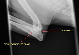
The most obvious radiographic feature in dogs with elbow dysplasia is development of osteophytes. These are normally first apparent on the dorsal margin of the anconeal process but eventually develop on all aspects of the joint (Figure 1). A fully flexed view of a well exposed radiograph is critical or the early signs will be missed. Unfortunately many affected dogs have no visible osteophytes. Only 70% of 263 dogs with confirmed medial coronoid disease had osteophytes on the dorsal margin of the anconeal process (1).

Is there another subtle radiographic sign that is useful?

Trochlear notch sclerosis is increased bone density below the region of the medial coronoid (Figure 1). Trochlear notch sclerosis (TNS) was identified in 86.7% of dogs with elbow dysplasia and medial coronoid disease. Another study described the sensitivity for correctly diagnosing medial coronoid disease using TNS as 91%-96% (2). This study also showed that the severity of the sclerosis significantly increased with increasing severity of coronoid disease grade as evaluated by CT.


Figure 1: Flexed lateral of the elbow. By fully flexing the elbow it allows visualisation of the osteophytes on the dorsal margin of the anconeal process. Trochlear notch sclerosis is characterised by increased bone density in the region adjacent to the medial coronoid.


Can dogs with elbow dysplasia have normal radiographs?

Unfortunately some dogs with medial coronoid disease can have unremarkable radiographs. Fitzpatrick reported 13 dogs (3%) of elbows had normal radiographs. Six of these had fragmentation, two had fissures and five had an intact MCP with significant chondromalacia (1).

Are older dogs easier to diagnose than younger dogs?

Diagnosis is not always easier in older dogs. A study comparing dogs with arthroscopically confirmed medial coronoid disease which were either less than or more than six years old showed no difference in the radiographic degree of osteoarthritis between the two groups.  Furthermore 20% of older dogs had erosions fissures or fragments despite a  0 IEWG score (3). The onset of lameness in the older dogs was frequently within months prior to presentation (irrespective of age of presentation) and arthroscopic fragment removal resulted in resolution or markedly reduced lameness.

If radiographs fail then what is the ‘gold’ standard?

Elbow CT is probably the most useful imaging modality for assessing elbows. It is the gold standard for diagnosing incomplete ossification of the humeral condyle and will detect most cases of medial coronoid disease (Figure 2). CT can also detect fragmentation of the medial coronoid process which may be missed arthroscopically (4). However differentiating fragmentation of the medial coronoid process from osteophytes can be challenging and similarly OCD cannot be easily differentiated from “kissing lesions” on the medial aspect of the humeral condyle [16]. Some non-displaced fissures appear to be solid bone on CT but most of these dogs will have an abnormal appearance to the medial coronoid process. It is recognised that cases with arthroscopically confirmed fissures can have normal CT. For these reasons CT should not be used to exclude arthroscopic treatment (4).

Figure 2: CT images demonstrating radial commissure fissures of the medial coronoid in a 16-month-old dog (left) and 7 year old dog (right). Both dogs had a history of lameness of less than 2 months and both dogs had dramatically reduced lameness (older dog) and resolved lameness (young dog) with fragment removal.

Do radiographic changes correlate with the severity of the disease?

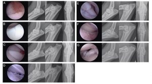
Unfortunately the radiographic severity in general does not correlate with the severity of disease (Figure 3). A recent abstract presentation on this topic suggested that if the osteophytes are greater than 5mm high then these are associated with more severe pathology identified on arthroscopy.

Figure 3: Arthroscopic images (left) with matched extended and flexed mediolateral radiographs and craniocaudal radiograph (left to right) demonstrating the spectrum of joint pathology identified. (A) 12-month-old male neutered German Short Haired pointer with moderate synovitis and modified Outerbridge grade 2 cartilage pathology at the tip of the MCP without overt fissure or fragment formation (left elbow); (B) 11- month-old male entire Rottweiler with fissure formation in the region of the radial incisure of the MCP (right elbow); (C) 18-month-old female neutered Labrador retriever with gross fragment formation at the radial incisure of the MCP (left elbow); (D) 28-month-old male neutered Golden Retriever with modified Outerbridge grade 3 pathology of both MCP and medial humeral condylar surface demonstrating typical linear striations in absence of gross fragmentation of the MCP (left elbow); (E) 15-month-old male entire English Springer Spaniel with modified Outerbridge grade 3–4 pathology of both MCP and medial humeral condylar surface including gross fragmentation of radial incisure of MCP with apparently undiseased cartilage cap (right elbow); (F) 60-month-old female neutered German Shepherd Dog with modified Outerbridge grade 5 pathology of both MCP and medial humeral condylar surface in absence of gross fragmentation of the MCP (right elbow); (G) 20-month-old male entire Golden Retriever with modified Outerbridge grade 5 pathology of both MCP and medial humeral condylar surface including gross fragmentation of radial incisure of MCP with apparently undiseased cartilage cap (left elbow). From Noel Fitzpatrick et.al. Radiographic and Arthroscopic Findings in the Elbow Joints of 263 Dogs with Medial Coronoid Disease Veterinary Surgery 38:213–223, 2009

References

1. Fitzpatrick, N., et al., Radiographic and Arthroscopic Findings in the Elbow Joints of 263 Dogs with Medial Coronoid Disease. Veterinary Surgery, 2009. 38(2): p. 213-223.

2. Draffan, D., et al., Radiographic analysis of trochlear notch sclerosis in the diagnosis of osteoarthritis secondary to medial coronoid disease. Veterinary and Comparative Orthopaedics and Traumatology, 2009. 22(1): p. 7-15.

3. Vermote, K.A., et al., Elbow lameness in dogs of six years and older. Veterinary and Comparative Orthopaedics and Traumatology, 2010. 23(1): p. 43-50.

4. Moores, A.P., L. Benigni, and C.R. Lamb, Computed Tomography Versus Arthroscopy for Detection of Canine Elbow Dysplasia Lesions. Veterinary Surgery, 2008. 37(4): p. 390-398.


For referring vets, please use our online referral form to submit a case enquiry.


Our Network

Animal Referral & Emergency network is the largest specialty and referral network in Australia, consisting of over 20 sites. With over 1,200 dedicated team members, including over 600 nurses and over 390 veterinarians (including specialists and registrars), we provide exceptional care for your pets. Count on us for expert medical attention and comprehensive veterinary services.