Avatar

Veterinary Clinical Conference - Connect 25 - Early Bird Ticket & Discount Now Available

Click here for more info (earn up to 11.5 CE points)
Search for AREN


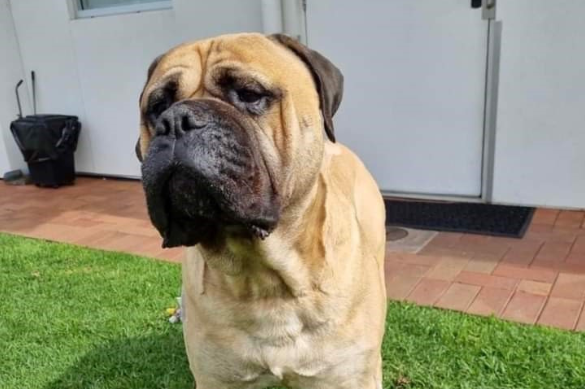
Meet Clyde, the Bull Mastiff that stole all of our hearts!

About Clyde and his condition

Meet Clyde, the 5 year old 65Kg Bull Mastiff who stole ALL our hearts! He was seen at his referring vet after a 7 day history of lethargy, tachypnoea and innapetence. Thoracic radiographs were performed and showed a fluid/soft tissue opacity that obscured the majority of the lung fields and cardiac silhouette. Thoracocentesis was performed and 1 litre of chyle like fluid removed. Fluid was submitted for evaluation and revealed a lymphocyte-rich effusion with Triglyceride levels higher than in serum. Clyde was referred to the AAERC Medicine Department for investigation.


Differential diagnoses for chylothorax included: Pericarditis/Cardiac disease, Intrathoracic mass eg thymoma, or cranial mediastinal neoplasia, Thoracic duct occlusion, Thromboembolic disease, Inflammatory disease and Idiopathic chylothorax.A diagnostic plan included thoracic ultrasound/echocardiogram with a view to performing a CT scan if there is no pathology evident on ultrasound. Right sided congestive heart failure can contribute to increased venous return and hence pleural effusion. A small degree of tricuspid valve disease was noted but not considered significant particularly in light of there being supporting clinical signs. No specific causes could be found on CT and therefore an idiopathic diagnosis was made. Ultrasound guided thoracocentesis was performed from the right thorax and additional chyle removed.


He was monitored over the following 12 months and after initial resolution developed recurrent episodes of coughing and dyspnoea with an acute life threatening episode of chylothorax where 4 litres of chyle was removed. Emergency thoracocentesis was performed on him in sternal recumbency. Using local anaesthetic and a small skin cut down, large 14 G over the needle catheters were inserted dorsal to the costochondral junction at the 8th IC space. Fluid was then removed using extension tubing attached to a 3 way stop cock and 50 ml syringe. If back pressure was encountered gently tilting him to the dependant side and manipulating the catheters trajectory into the chest allowed for maximal flow. Medical management including low fat dietary modification helps and occasionally resolve spontaneously but idiopathic forms often require thoracic duct ligation to stop the effusion. Chronic effusion has also been shown to increases the risk of fibrosing pleuritis which in itself causes dyspnoea, hence the owner’s decision to do surgery


Pre-operative CT lymphangiography can help visualize the various branches of the thoracic duct. This may be performed by injecting Iohexol into either the popliteal, mesenteric lymph nodes or an iodine contrast agent subcutaneously around the anus. Intraoperative mesenteric lymphangiography and fluoroscopy has also been described. A practical technique and one used in this case involved diluted methylene blue injected into a lymphatic or intranodally. This is very helpful in identifying all branches of the thoracic duct helping to ensure complete ligation.  

Clyde was positioned in left lateral recumbency and a right sided 9th intercostal thoracotomy performed after he was placed on a ventilator. A local block was carried out at the two ribs cranial and caudal to the thoracotomy using Bupivacaine. A further 200 ml of chyle was suctioned and the chest lavaged with saline. The pleura was thickened on its reflection at the mediastinum. The aorta, sympathetic chain and azygos vein identified. A para costal incision was made and after exteriorising a loop of duodenum an attempt at catheterisation of an efferent lymphatic for lymphangiography was performed.


The lymphatics were very friable and after two attempts at catheterization this was abandoned. A lymph node at the ileocolic mesentery was then injected with 0.5 ml of Methalyne blue and then 5 minutes later. The thoracic duct and its associated branches could be now be clearly visualised in the right mediastinum. Thee large ducts could be seen in the peri aortic tissue and three 10mm liga clips were applied. A 2 cm incision was made dorsal to the aorta and a number of smaller ancillary branches were seen. An encircling 2/0 Prolene ligature was passed through the mediastinum and used to ligate these en bloc.A right sided subtotal pericardectomy was performed by making a T shaped incision ventral to the phrenic nerve and extending to the apex of the heart thereby removing a triangular area of thickened pericardium. The chest and abdomen were lavaged with copious amounts of warm saline and a chest drain placed. Layered closure of the thoracotomy and para costal incision was performed.


After surgery, Clyde was closely monitoring by a dedicated hospital team overnight, then spent a few days with our Emergency and Critical Care Team. Clyde has done extremely well post operatively and at 3 months has no further symptoms.

  • Useful facts: (Reference: Fossum) 
  • Chyle is fatty lymphatic fluid circulated from the intestine to the cranial vena cava. 
  • Any factors that increase flow or decrease drainage may dilate the lymphatics Causing lymphanioectasia. 
  • Any disease that increased systemic venous pressure can cause chylothorax 
  • A history of coughing is often the first symptom then later followed by dyspnoea. 
  • Radiographs are a useful diagnostic tool. Chyle is typically white or pink and occasionally red in colour.( Pink milkshake) 
  • Suspected chylous effusions should be submitted in EDTA. 
  • Diagnostic characteristics include: Triglyceride content (Higher than simultaneously collected serum) 
  • Cytology (Predominant cell type is lymphocytes or non-degenerative neutrophils) 
  • CT lymphangiography can be useful but not essential in the diagnosis and treatment 
  • If the underlying disease is diagnosed and treated appropriately many cases resolve  
  • Surgery is warranted where the underlying cause is unknown (idiopathic) and effusions become chronic. 
  • Low fat diets are non-curative. Surgical treatment involves:  
    TD ligation and pericardectomy* (commonly performed, often done simultaneously)
  •  Success rate with TDL and subtotal pericardectomy is in the region of 80% Cisterna Chyli ablation and less commonly TD glue embolization and active pleuro peritoneal shunting are also described.  
  • Pericardectomy is thought to lower right –sided venous pressure normalizing venous pressure and lymphatic flow.  

Our Network

Animal Referral & Emergency network is the largest specialty and referral network in Australia, consisting of over 20 sites. With over 1,200 dedicated team members, including over 600 nurses and over 390 veterinarians (including specialists and registrars), we provide exceptional care for your pets. Count on us for expert medical attention and comprehensive veterinary services.